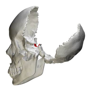
The sphenoid sinus is an unique and intricate structure that is located posteriorly to the other paranasal sinuses near the base of the skull. The sphenoid sinus’s pattern of pneumatization and the juxtaposition to various other neurovascular structures exhibit notable inter-individual differences, making them an intriguing topic for research in the area of anatomy. Notably, sinus expansion remains throughout life and reaches its peak only after puberty, which challenges the research of this structure.
The diaphysis of the sphenoid bone contains the sphenoidal sinuses, also known as pneumatic cavities, which are lined with mucous membrane. It is well known that the morphology of the sphenoid sinus exhibits significant variation with regard to its size, shape, number of septa, and level of air fill. These variances might present difficulties in clinical and surgical applications, highlighting the need of having a complete grasp of the sphenoid sinus’ anatomy. Previous research demonstrates the variability of the sphenoid sinus.
The Endoscopic Endonasal Transsphenoidal Approach (EETA)
The endoscopic endonasal transsphenoidal approach (EETA) is a surgical modality that has gained widespread acceptance for the treatment of pituitary adenomas and other pathologies of the skull base. This approach is preferred due to its remarkable safety profile and a low incidence of complications, which can be further mitigated by utilizing precise anatomical knowledge. The procedure involves accessing the lesion via the nostrils and sphenoid sinus, using endoscopes and other specialized surgical instruments to perform the resection. Significantly, the EETA technique offers a direct, less invasive route to the target lesion in comparison to traditional open surgical procedures. This technique has been shown to deliver favorable outcomes, including reduced blood loss, shorter hospital stays, and faster recovery times, thereby benefiting both the patient and the healthcare system. The EETA has become an expedient technique, used during surgical operations for most parasellar and intrasellar tumor procedures. In EETA, the morphological characteristics of the sella turcica are noteworthy. The size, form, and pneumatization changes of the sella turcica, which is used in this surgical technique to reach the pituitary gland, might affect the outcome of the treatment. For preoperative planning and intraoperative navigation, an understanding of sella turcica shape is vital since it affects surgical access and visualization. Different conditions may be linked to various sella turcica anomalies, which can help with diagnosis and therapy. To improve surgical results and patient care, it is important to highlight the clinical significance of this association in EETA.
The transnasal approach, as compared to open craniotomy, is considered a less invasive technique resulting in lower morbidity and mortality rates. Endoscopic visualization of hard-to-reach areas is facilitated by the technique and the EETA has emerged as a preferred approach for most parasellar and intrasellar tumor procedures. In the pediatric population, EETA is preferred because it is less traumatic, has a shortened recovery time, and does not impact the anatomical and functional integrity of the skull, nor hinder growth in young patients. Sphenoid sinus morphology exhibits significant inter-individual variations, with differing size, pneumatization, and septation patterns leading to variations in sphenoid sinus segmentation.
Studying sella turcica shape and sphenoid pneumatization types has therapeutic implications for many different medical specialties. Understanding these characteristics from a radiological perspective makes it easier to accurately interpret the imaging findings and discern abnormalities and diseases. Understanding sella turcica morphology in neurosurgery is essential for surgical planning, especially when it comes to the pituitary gland, lowering risks and enhancing outcomes.
Pituitary Adenoma
Adenomas of the pituitary and other endocrinological conditions can be connected to anomalies of the sella turcica, making early detection and therapy attainable. The anthropological and forensic importance of these investigations extends beyond medicine, giving insights on human evolution and demographic distinctions. Additionally, sella turcica measurements in orthodontics affect how to manage craniofacial problems.
The applicability of understanding sphenoid pneumatization variations and sella turcica morphology across numerous dental disciplines are what give this research its dental clinical value. It offers insights on craniofacial development in orthodontics, assisting with individualized treatment regimens. Surgical planning for operations in the midface is advantageous for oral and maxillofacial surgery
The placement and design of dental prostheses are improved, which enhances prosthodontics. Additionally, it could provide information on how to treat temporomandibular joint disorders. Additionally assisted is accurate picture interpretation in dental radiology. Overall, this understanding improves dental therapy, resulting in greater healing and patient wellbeing.
Several previous studies have highlighted the importance of careful preoperative planning and imaging to ensure optimal surgical outcomes. The anatomical structure of the sphenoid sinus and variations in bone anatomy in this region can be clearly seen through radiological examination. Despite the extensive literature on the sphenoid sinus and sella turcica, the relationship between the type of pneumatization of the sphenoid sinus and the sella turcica has not received sufficient attention. Therefore, this study seeks to fill this knowledge gap by investigating the potential correlation between different types of pneumatization of the sphenoid sinus and sella turcica.
Author: Prof. Dr. Dian Agustin Wahjuningrum, drg., Sp.KG. Subsp, KE(K)
More detailed information from this research can be seen in our article at: https://peerj.com/articles/16623/
Mehmet Emin Dogan, Sedef Kotanlı, Yasemin Yavuz, Dian Agustin Wahjuningrum and Ajinkya M. Pawar. [2023] Computed tomography-based assessment of sphenoid sinus and sella turcica pneumatization analysis: a retrospective study.