
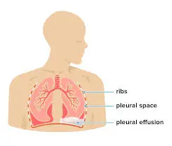
Hepatic Hydrothorax is an accumulation of transudative pleural effusion (generally ≥500 ml) in patients with chronic liver disease and portal hypertension, after excluding other etiologies such as heart, lung, kidney disorders and so on. Pleural fluid comes from capillaries on the surface of the parietal pleura which move based on the Starling force and are reabsorbed by lymphatic stomata on the surface of the parietal pleura. Especially in the mediastinum and diaphragm regions so as to maintain the volume of pleural fluid within the normal range of 0.1 ml/kg to 0.3 ml/kg. Fluid can also enter the pleural cavity from the pulmonary interstitium through the visceral pleura. Or from the peritoneal cavity through small holes in the diaphragm at a speed of 0.5 ml/hour. Hepatic hydrothorax occurs due to direct movement of fluid from the peritoneum to the pleural cavity through a diaphragm defect. This diaphragmatic defect usually measures <1 cm and generally occurs on the right side (85%), with 13% occurring on the left side and 2% occurring bilaterally.
Symptoms of Hepatic Hydrothorax
Respiratory symptoms in hepatic hydrothorax vary, ranging from asymptomatic, coughing, pleuritic chest pain, shortness of breath, to life-threatening respiratory distress. Most effusions were mild to moderate, and only 6% had massive effusions that filled more than half of the hemithorax. Some patients have no symptoms and are incidental findings on radiological examination, but the majority of cases have symptoms in the form of: dyspnea at rest (34%), cough (22%), nausea (11%) and pleuritic chest pain (8%). In patients with HH there are signs and symptoms of hepatic cirrhosis and portal hypertension. For example ascites, spider naevi, asterixis, hepatosplenomegaly, caput medusa, and hepatic encephalopathy.
In patients with hepatic hydrothorax with signs and symptoms of fever, pleuritic chest pain, or encephalopathy, it is necessary to consider the possibility of a cavum pleura infection, such as spontaneous bacterial empyema (SBEM) (also called spontaneous bacterial pleuritis [SBPL]). SBEM or SBPL is a complication that can occur due to the migration of a bacterial infection in the peritoneum to the cavum pleura or a bacterial invasion through pleural chest tubes, catheters, or other instruments.
Diagnosis of Hepatic Hydrothorax
Hepatic hydrothorax is a diagnosis of exclusion. Therefore, other etiologies of pleural effusion, such as cardiac, pulmonary, pleural disorders, and malignancy, must be ruled out first. Supportive examinations, such as echocardiography and brain natriuretic peptide (BNP) blood tests, can be performed to evaluate cardiac function. CT of the thorax can exclude other causes, such as mediastinal malignancy, and lung or pleural lesions, and abdominal ultrasound can be performed to evaluate the hepatic mass, ascites, and hepatic and portal venous flow.
The diagnosis of hepatic hydrothorax is based on clinical manifestations, radiologic features, and thoracocentesis to exclude other etiologies such as infection (spontaneous bacterial peritonitis [SBP], tuberculosis), malignancy (lymphoma, adenocarcinoma) and chylothorax. The management of hepatic hydrothorax is complex, involving a multidisciplinary approach. The management strategy includes a stepwise approach of one or more of the following five methods: 1) Reducing ascitic fluid production; 2) Preventing fluid transfer to the pleural space; 3) Fluid drainage from the pleural cavity; 4) Pleurodesis (obliteration of the pleural cavity); 5) Liver transplantation.
The conclusion of this article is that the management of Hepatic Hydrothorax is quite complex and requires a multidisciplinary approach by considering the patient’s clinical condition, the risk of complications that may occur and the availability of health personnel and facilities. Correct diagnosis and therapy are the keys to successful management of Hepatic Hydrothorax. Apart from looking at clinical manifestations and radiological examinations, pleural fluid analysis is needed to determine the diagnosis and assess complications in Hepatic Hydrothorax.
Detailed information from this research can be seen in the following article link: https://www.kjg.or.kr/journal/view.html?uid=5992&vmd=Full&
Author: Amie Vidyani, dr., Sp.PD.